What Your Stool Color Means: A Complete Medical Guide
Most people glance and flush. But the color of your stool is one of the most information-dense, immediately available windows into your gastrointestinal health — and knowing how to read it takes minutes to learn and can matter enormously.
Why Stool Has Color in the First Place
Normal stool is brown — a fact so unremarkable that most people never stop to wonder why. The answer lies in one of the most biochemically fascinating waste-processing systems in the body: bile.
Red blood cells have a lifespan of approximately 120 days. When they reach the end of their cycle, they are broken down in the spleen and liver. The iron-containing hemoglobin molecule is stripped apart, and the porphyrin ring left behind is converted into bilirubin — a yellow-orange compound. The liver conjugates bilirubin to make it water-soluble and secretes it into bile, the digestive fluid produced by the liver and stored in the gallbladder. When you eat a fat-containing meal, the gallbladder contracts and releases bile into the small intestine to help emulsify dietary fats.
As bilirubin travels through the intestinal tract, bacteria in the colon chemically transform it through a series of reduction reactions, ultimately converting it to a compound called stercobilin. Stercobilin is the pigment responsible for the characteristic brown color of normal stool. The more bilirubin that makes it into the stool (which correlates with how much bile is produced and how much reaches the colon intact), and the degree of bacterial conversion, determines the shade — from light tan to very dark brown.
This mechanism means that stool color is a direct readout of three intersecting systems: liver and gallbladder function (how much bile is produced and released), intestinal transit time (how long contents stay in the colon and are exposed to bacteria), and diet (certain pigmented foods pass through intact and temporarily shift color). Understanding which system is responsible for a color change allows you to interpret it correctly.
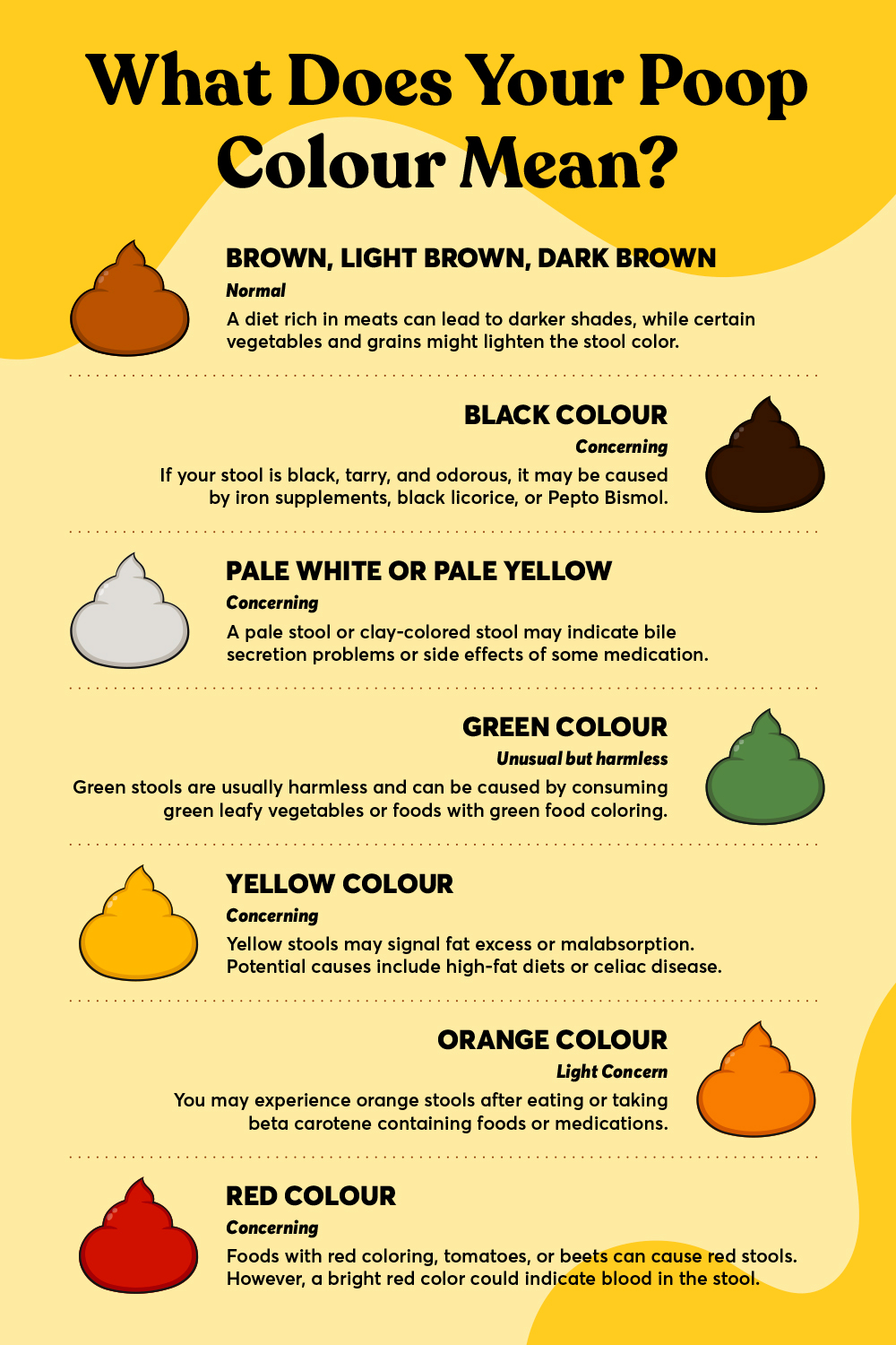
Brown: The Healthy Baseline
The full spectrum of healthy stool color runs from light tan and khaki through medium brown to a rich chocolate brown. All shades within this range are normal and are simply reflections of subtle variations in the amount of bile excreted, how long transit takes that day, and what was eaten. A tan, putty-colored stool after a very low-fat meal (which triggers minimal bile release) followed by a darker brown stool after a fatty dinner is completely normal variation within the same individual on the same day.
The consistency of stool — its Bristol Stool Scale type — carries at least as much diagnostic information as color in most circumstances. A dark brown Type 1 (hard pellets) tells a different story than a dark brown Type 4 (smooth sausage). When evaluating any stool, always consider color and consistency together.
Yellow Stool: Fat Malabsorption and Speed
Yellow stool occupies an important clinical territory because it can indicate either a completely benign cause or a significant malabsorption problem.
The Benign Cause: Fast Transit
If stool moves through the colon faster than usual — triggered by a high-fiber meal, stress, a gastroenteritis episode, or simply natural variation — it has less time to be processed by colonic bacteria and less time for stercobilin to develop fully. The result is yellow, mushy stool that may have a more acidic odor than usual. This kind of yellow stool is often accompanied by a Bristol Type 6 or 7 consistency, resolves within a day or two, and requires no medical attention if it coincides with a clear dietary or stress trigger.
The More Serious Cause: Fat Malabsorption (Steatorrhea)
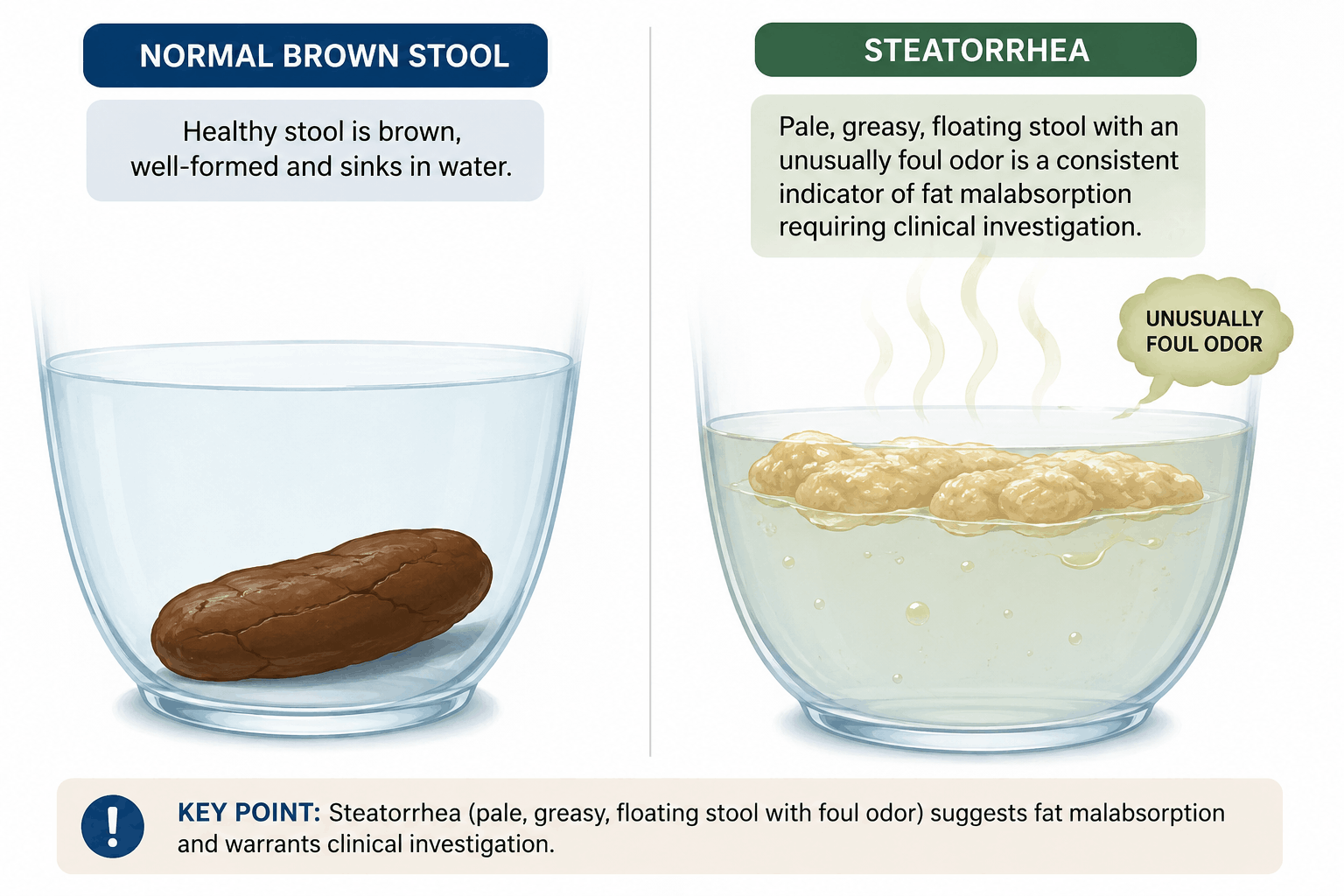
Yellow, greasy, unusually foul-smelling stool that floats persistently is a distinct clinical sign called steatorrhea — the presence of excess fat in the stool. When fat is not properly digested and absorbed in the small intestine, it reaches the colon intact, producing stool that is pale yellow to orange-yellow, oily in texture, difficult to flush, and markedly malodorous due to bacterial fermentation of undigested lipids.
Steatorrhea is a diagnostic red flag for malabsorption disorders and can indicate:
- Exocrine pancreatic insufficiency (EPI): The pancreas produces insufficient lipase (fat-digesting enzyme). Causes include chronic pancreatitis, cystic fibrosis, and pancreatic cancer. EPI is underdiagnosed — studies suggest it affects 5–10% of the general population, with rates dramatically higher in people with a history of heavy alcohol use or chronic pancreatitis.
- Celiac disease: Villous atrophy in the small intestine reduces the absorptive surface area for fats, bile acids, and fat-soluble vitamins (A, D, E, K). Read more in our article on celiac disease.
- Bile acid malabsorption (BAM): Bile acids are normally reabsorbed in the terminal ileum and recycled; if this reabsorption is disrupted (by Crohn's disease affecting the ileum, prior ileal resection, or primary BAM), fat digestion is impaired.
- Short bowel syndrome: Following significant intestinal resection, the reduced absorptive length produces persistent steatorrhea.
- Giardia infection: The parasite Giardia lamblia adheres to the intestinal wall and directly impairs fat absorption, producing yellow, greasy, foul-smelling stools. A hallmark of traveler's diarrhea in certain regions.
Persistent yellow, fatty stools lasting more than a week — particularly if accompanied by weight loss, abdominal pain, or fatigue — require a physician evaluation. Diagnosis typically involves stool fat analysis (72-hour fecal fat collection), fecal elastase testing for pancreatic function, and serology for celiac disease.
Green Stool: Diet, Speed, and Bile
Green stool is among the most common color changes people worry about, and in the vast majority of cases, it is entirely harmless. That said, the mechanism matters for interpretation.
Dietary Green
The most obvious cause: eating large quantities of green vegetables — spinach, kale, broccoli, arugula, collard greens — can temporarily produce green-tinted stool. Chlorophyll, the green pigment in plants, is not fully broken down during digestion in some individuals and can persist as a mild colorant. Iron supplements and certain multivitamins also commonly produce green or dark green stools, which is harmless.
Artificially colored foods are another frequent culprit that people often overlook. Green food dye (found in certain sports drinks, artificially colored candy, green-colored desserts, and heavily colored cereals) will pass through and produce bright green stools that cause unnecessary alarm. If you've recently consumed any heavily dyed food or drink, that's almost certainly the explanation.
Fast Transit Green
The second mechanism is transit-time related. Bile starts as yellow-green in color when it enters the small intestine. As it travels through the colon and is progressively converted by bacteria, it transitions from green-yellow to tan to brown. If transit is fast enough — due to diarrhea, IBS-D flares, acute gastroenteritis, or anxiety-driven colonic acceleration — bile doesn't have time to complete this conversion. The result is bright green or greenish-yellow stool that typically appears alongside loose, Bristol Type 5–7 stools. It is the color you often see during a stomach bug or a very stress-heavy day.
When Green Stool Is Worth Noting
If bright green stool appears without a clear dietary explanation, persists more than a week, is accompanied by urgency or cramping, or is associated with significant abdominal pain, it warrants a GP visit. Clostridioides difficile infection can occasionally produce green-tinged stool (though it more typically produces yellow), and certain bacterial GI infections can produce green stools as part of an acute illness that benefits from treatment rather than watchful waiting.
Black Stool: Upper GI Bleeding Until Proven Otherwise
Black, tarry stool — clinically called melena — is one of the most important color changes to recognize because it is frequently a sign of serious upper gastrointestinal bleeding and requires same-day medical evaluation.
What Melena Looks Like and Why
Melena is not just dark brown stool. It is jet-black, very sticky (described as tar-like), has a distinctive extremely foul, metallic smell unlike normal stool, and is often semi-liquid. The characteristic appearance results from a specific biochemical process: when blood bleeds into the upper GI tract (esophagus, stomach, or upper small intestine), gastric acid and digestive enzymes chemically alter the hemoglobin in the red blood cells, breaking it down into oxidized hemoglobin compounds — chiefly hematin — that are intensely black. As this altered blood mixes with stool and passes through the intestine, it produces the characteristic tarry consistency and intense odor.
A crude but useful clinical rule: blood entering the GI tract from anywhere at or above the ligament of Treitz (the junction of the duodenum and jejunum) will typically produce melena. Blood entering below this point — and particularly below the sigmoid colon — will typically produce bright red or maroon blood in the stool (hematochezia). This is the anatomical basis of the color-to-location inference described in our article on rectal bleeding.
Common Causes of True Melena
- Peptic ulcer disease: The most common cause of melena worldwide. Gastric or duodenal ulcers that erode into a blood vessel. Major risk factors: Helicobacter pylori infection, chronic NSAID use (ibuprofen, naproxen, aspirin), and heavy alcohol consumption.
- Esophageal varices: Dilated veins in the esophagus that form as a consequence of portal hypertension (elevated pressure in the portal venous system, most commonly due to cirrhosis). Variceal hemorrhage is a medical emergency; it can produce massive, rapid melena accompanied by hematemesis (vomiting blood) and hemodynamic instability.
- Mallory-Weiss tear: A longitudinal tear at the gastroesophageal junction caused by forceful or repeated vomiting or retching. Often produces self-limiting melena in the context of an acute vomiting episode.
- Gastric cancer: Upper GI malignancies can bleed intermittently for weeks or months before being diagnosed. Unexplained, persistent melena in someone over 45 should always prompt endoscopic evaluation.
- Angiodysplasia: Abnormal, fragile blood vessel formations in the GI mucosa that bleed without the dramatic presentation of other causes; more common in older adults.
The Iron Supplement Confound
There is one common and completely benign cause of very dark or black stool that is extremely important to know about: iron supplements. Ferrous sulfate and other oral iron formulations reliably produce dark green to black stools in most people who take them. The stool tends to be formed (not tarry), does not have the characteristic melena odor, and is directly temporally associated with starting supplementation. If you're taking iron and develop black stool, confirm with your prescribing clinician that this is expected for your formulation before attributing it to a GI bleed. Bismuth subsalicylate (Pepto-Bismol) also produces harmless black stools by reacting with hydrogen sulfide in the gut. These are important facts to know when reviewing stool logs.
Bright Red or Maroon Stool: Lower GI Bleeding
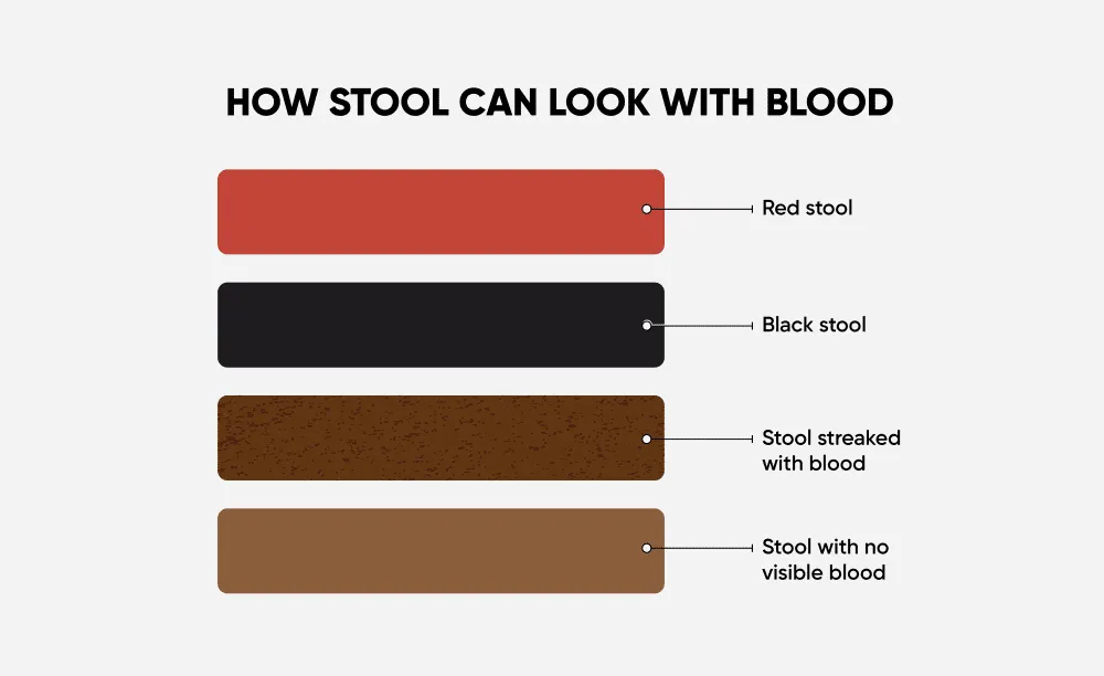
Bright red blood in or on the stool — hematochezia — indicates bleeding from the lower gastrointestinal tract: the colon, rectum, or anus. Unlike melena, the blood has not had time to be chemically altered by gastric acid, so it retains its red color. Localization based on color:
- Blood only on the toilet paper or coating the outside of a normal stool: Almost always from the anorectal area — hemorrhoids or an anal fissure. Very common, often associated with straining, constipation, or hard stools.
- Blood mixed through the stool: Suggests a source higher up — in the rectum or sigmoid colon. Can indicate hemorrhoids, rectal polyps, rectal cancer, or proctitis.
- Maroon, dark red blood mixed with stool, possibly clotted: Suggests a source in the more proximal colon. Can indicate diverticular bleeding (the most common cause of significant acute lower GI bleeding in adults over 50), colitis (infectious, ischemic, or inflammatory bowel disease), or colon cancer.
- Massive bright red bleeding with clots, accompanied by dizziness or rapid heart rate: A lower GI hemorrhage — requires emergency evaluation.
A single episode of small-volume rectal bleeding in a young adult with a history of constipation and straining is very likely a hemorrhoid or fissure. However, any rectal bleeding in a person over 45, rectal bleeding that persists more than 2–3 weeks, or rectal bleeding accompanied by a change in bowel habits, weight loss, or abdominal pain should be evaluated by a physician promptly. These are potential red-flag symptoms for colorectal cancer, which is highly treatable when caught early.
White, Pale Grey, or Clay-Colored Stool: Bile Duct Obstruction
Pale, chalky white, grey, or clay-colored stool is among the most clinically significant color changes you can observe, because it indicates a near-complete absence of bile reaching the intestine — a condition called acholic stool. Without stercobilin from bile to pigment it, stool loses its brown color entirely.
Normal pale stool immediately after a low-fat meal (which triggers minimal bile release) can occasionally appear lighter than usual, but truly white or clay-colored stool means one of two things:
- Bile duct obstruction: A blockage preventing bile from reaching the intestine. Common causes include gallstones blocking the common bile duct (choledocholithiasis), cancer of the pancreatic head compressing the bile duct (a notoriously "silent" early symptom), primary sclerosing cholangitis, or benign bile duct stricture after surgical or procedural injury.
- Severe liver disease: If the liver's ability to produce and conjugate bilirubin is severely impaired (acute hepatitis, acute liver failure, advanced cirrhosis), bile output to the intestine drops dramatically, producing pale stools.
Acholic stool is almost never a transient dietary variation. It is frequently accompanied by jaundice (yellowing of the skin and sclera) and dark urine — the yellow-brown discoloration of urine from bilirubin that the kidneys are excreting because it cannot exit via bile. This classic triad (pale stool, dark urine, jaundice) is clinically diagnostic of obstructive jaundice and requires urgent medical evaluation. It is one of the presentations of pancreatic cancer, which has a dramatically better prognosis when identified at an early, operable stage.
Note for parents: Pale, chalky stool in newborns and young infants requires immediate medical attention, as it can be a sign of biliary atresia — a rare but serious condition in which the bile ducts are absent or blocked from birth. Early surgical intervention (Kasai procedure) is time-sensitive and dramatically improves outcomes.
Orange Stool: Usually Diet, Rarely Something More
Orange stool is overwhelmingly dietary in origin and rarely indicates anything concerning. The most common cause is a large intake of beta-carotene — the orange pigment found in carrots, sweet potatoes, butternut squash, and pumpkin. Beta-carotene is a fat-soluble precursor to vitamin A that, when consumed in large amounts, can temporarily pigment both the skin (carotenodermia, particularly visible on the palms and soles) and the stool. The effect is dose-dependent and resolves completely when intake is reduced.
Orange food dyes (in sports drinks, artificially colored snacks, and certain medications) can also produce orange stools. Rifampin, an antibiotic used to treat tuberculosis and certain other infections, is famous for turning urine, tears, sweat, and sometimes stool a distinctive orange-red color. This is expected and harmless with the medication.
Less commonly, orange stool can indicate bile salt malabsorption, in which bile salts that are normally reconstituted in the terminal ileum instead reach the colon and alter stool pigmentation. If orange stools appear without dietary explanation and persist, particularly alongside diarrhea and fat malabsorption signs, a clinical review is warranted.
Why Stool Color Tracking Matters Over Time
A single observation of stool color provides context but rarely delivers a conclusion. What provides genuine medical value is a documented pattern over time. The difference between "I've noticed my stool has been paler recently" and "I have a 23-day log showing a consistent shift from medium brown to pale tan, with fatty texture noted on 11 of those days" is the difference between a vague complaint and actionable clinical data.
This is precisely the function of color tracking in Happy Poop. Each log entry allows you to record your stool's color from a standardized palette — capturing not just a subjective impression, but a timestamped, categorized data point that you can chart over weeks and months. The Dashboard shows color distribution over any selected time period, making it immediately clear whether a color change is a one-off event or a sustained pattern.
When you see your gastroenterologist or GP and they ask "how long has your stool been this color?", a precise answer — backed by a data export showing the color history — is clinically far more valuable than "a few weeks, maybe?" It shortens the diagnostic process, helps distinguish genuine pathology from dietary variation, and reduces the chance of a concerning sign being dismissed as benign.
Download Happy Poop and start logging today:






Quick Reference: Stool Colors at a Glance
The following is a summary reference. Always correlate color with consistency, frequency, accompanying symptoms, diet, and medications before drawing conclusions.
- Light tan to dark brown: Normal range. No action required.
- Yellow, formed: Usually fast transit or high-fat meal. Persistent floating, greasy yellow stool → fat malabsorption workup.
- Green, formed: Diet (leafy greens, iron, food dye) or fast transit. Persistent green with diarrhea and pain → clinical review.
- Black, tarry, foul-smelling: Upper GI bleeding until proven otherwise. Seek same-day medical evaluation. (Exception: iron supplements, bismuth products.)
- Bright red blood on/in stool: Lower GI source. Isolated small amount in young adult with constipation → likely hemorrhoid. Any age 45+, persistent, or with changed habits → urgent clinical review.
- Pale grey / white / clay: Bile duct obstruction or severe liver disease. Urgent medical evaluation — do not wait.
- Orange: Almost always beta-carotene or food dye. Persistent without dietary cause → clinical review.
Sources & Medical References: